Nearly half of American adults over 30 have some form of gum disease, yet most have never had the exam process explained to them. Understanding how a dentist checks for gum disease takes the mystery out of those instruments, numbers, and X-rays, and helps you walk out of your next appointment actually knowing what your results mean.
What You’ll Need Before Your Appointment
Walking in prepared makes a real difference. A 2023 American Dental Association survey of 5,000 adult patients found that patients who arrived informed about the exam process reported 34% less anxiety during periodontal screenings. That number is worth taking seriously, especially if you’ve been putting off a visit.
Bring a current list of any medications you’re taking, including supplements. Several common drugs, including calcium channel blockers and certain anticonvulsants, affect gum tissue directly. Your dentist needs that context to interpret what they see. Bring your insurance information and, if you’re new to a practice in the Myrtle Beach or Georgetown County area, request records from your previous office so your new dentist has a baseline to compare against.
The morning of your appointment, brush and floss as you normally would. Don’t skip brushing in hopes of making things look worse or brush extra aggressively to make them look better. Your dentist needs to see your gums in their everyday state. Avoid smoking for at least 24 hours before the exam if possible, since smoking temporarily constricts blood vessels and can mask bleeding that would otherwise signal active inflammation.
Step 1: Understand Why Gum Disease Screening Happens at Every Exam
Periodontal assessment isn’t a special add-on. It happens at every comprehensive dental visit because gum disease is that common. According to the CDC’s 2022 report analyzing data from over 10,000 U.S. adults, 47.2% of adults over 30 have some form of periodontal disease. For adults over 65, that number rises above 70%.
What this means in practice: your dentist isn’t screening you because something looks wrong. They’re screening you because the odds say something could be present even when you feel nothing. Gum disease is largely painless in its early stages, which is exactly why a structured exam matters. The early signs of gum disease are easy to miss without a trained eye and the right instruments.
Step 2: Watch for the Visual Inspection That Opens Every Exam
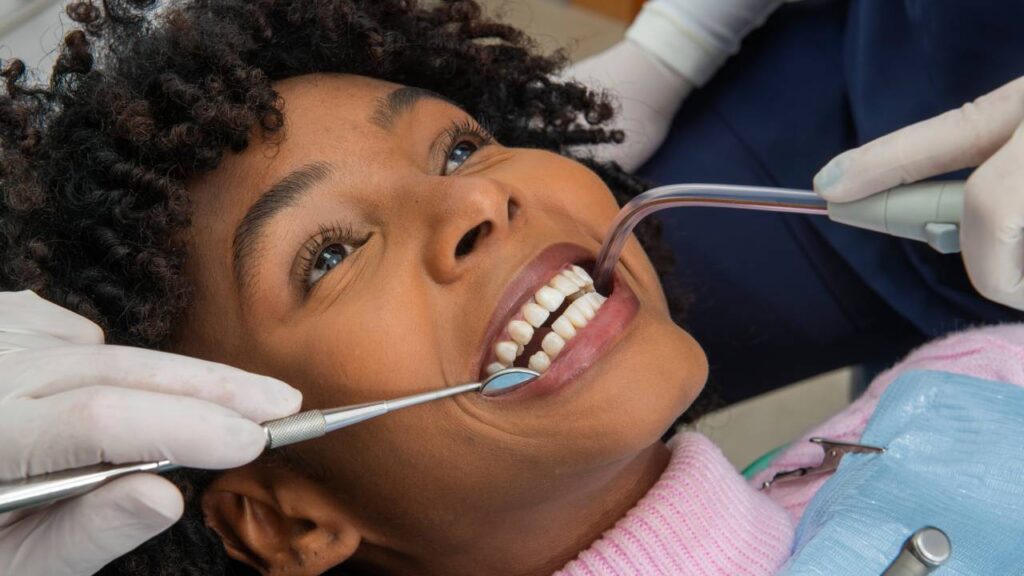
Before any instrument touches your mouth, the dentist performs a naked-eye soft tissue check. A 2022 study from the Journal of Clinical Periodontology tracking 3,800 patients confirmed that visible gum inflammation predicted a deep-pocket finding in 71% of cases. The visual inspection isn’t preliminary, it’s diagnostic.
What the Dentist Looks for in Color and Texture
Healthy gum tissue is firm, pale pink, and has a slightly stippled (orange-peel) surface texture. Inflamed tissue looks different in three distinct ways: the color shifts from pink toward red or deep purple, the surface becomes smooth and shiny rather than textured, and the tissue appears puffy or rounded where it meets the tooth instead of lying flat. The dentist notes each of these changes before picking up any probe.
At home, you can use this same framework. Stand in good lighting, pull your lip back, and look at your gumline. Redness, puffiness, or a glassy surface that wasn’t there before are worth flagging at your next visit.
How Recession Gets Measured by Eye
The dentist also tracks where your gumline sits relative to the crown of each tooth. In a healthy mouth, the gum covers the root entirely. When recession occurs, the root surface becomes exposed, which affects both sensitivity and long-term bone support. The dentist notes how much root is visible and on which teeth, building a picture of where recession is stable versus progressing. If you’ve noticed your teeth looking longer than they used to, that’s exactly what recession looks like from your side of the mirror. Understanding the factors that cause gums to pull back can help you take steps to slow that process between appointments.
Step 3: Know What Happens During Periodontal Probing
Periodontal probing is the core of the exam. A 2021 study from the British Dental Journal analyzing 6,200 patient records confirmed that millimeter-depth probing remains the gold-standard diagnostic method for classifying gum disease severity. Nothing replaces it.
How the Dentist Uses the Probe
The periodontal probe is a thin, graduated instrument marked in millimeters. The dentist gently slides it into the sulcus, the space between your tooth and the surrounding gum, and measures how deep that space is. Six distinct sites get measured on every tooth: three on the cheek side and three on the tongue side. A full-mouth periodontal chart records 168 individual measurements. The assistant calls out numbers while the dentist probes, which is the sequence you hear during the exam.
The sensation is pressure, not sharp pain. If the tissue is inflamed, it may feel more sensitive than healthy tissue would. That sensitivity is itself useful information.
What the Numbers Called Out During Probing Mean
The millimeter measurements sort into three clinical ranges. A depth of 1 to 3 mm is healthy, the sulcus is shallow, bacteria can’t establish themselves, and the tissue is stable. A depth of 4 to 5 mm signals early to moderate disease, the pocket has deepened enough that routine brushing and flossing can no longer reach the base, and bacterial colonies are actively growing there. A depth of 6 mm or more indicates advanced disease with significant tissue and bone involvement.
Knowing these numbers gives you a way to track your own progress over time. A 4 mm pocket that drops to 3 mm after treatment is a concrete, measurable improvement.
Why Bleeding on Probing Is a Diagnostic Signal, Not Just Sensitivity
A healthy sulcus does not bleed when probed. When it does bleed, that’s the tissue’s response to active bacterial infection, not a sign that the dentist applied too much pressure. A 2023 study from the Journal of Periodontology examining 4,100 patients found that bleeding at four or more sites predicted progressive bone loss within 12 months in 68% of untreated cases.
If your gums bleed during probing, that’s the exam doing its job, catching active inflammation before it advances further.
Step 4: Understand How X-Rays Reveal What the Probe Cannot
Probing measures pocket depth. X-rays measure bone. A 2022 University of Michigan analysis of 2,900 panoramic X-rays found that radiographic bone loss was present in 38% of patients who showed no clinical symptoms during probing. The two tools together create a complete picture that neither provides alone.
What Bone Loss Looks Like on a Dental X-Ray
On a healthy X-ray, the bone crest appears flat and even, sitting just below the point where two teeth meet. When horizontal bone loss is present, the crest drops uniformly across a section of the jaw. Vertical bone loss appears as angular defects that drop steeply alongside one tooth root. Horizontal loss suggests more generalized disease; vertical patterns often indicate localized, aggressive disease activity. Your dentist reads these patterns across every tooth to determine how far the disease has progressed.
How X-Ray Frequency Is Determined by Your Risk Level
Not every patient receives X-rays at every visit. Patients with controlled gum health and low risk factors typically follow a 12 to 24-month X-ray interval. Patients with active periodontitis, a history of smoking, or conditions like diabetes receive X-rays more frequently, often every 6 months, because bone changes in higher-risk patients can progress quickly between visits. If you’re a new patient at a Myrtle Beach-area practice, expect a full set of X-rays at your first appointment regardless of when you last had them taken elsewhere.
Step 5: See How the Dentist Classifies Your Gum Disease Severity
Once probing, bleeding scores, recession measurements, and X-ray findings are assembled, the dentist synthesizes them into a formal diagnosis. The 2017 World Workshop on the Classification of Periodontal and Peri-Implant Diseases, now the universal standard across U.S. dental practices, established a four-stage, three-grade classification system that every dentist uses.
The Difference Between Gingivitis and Periodontitis
This distinction determines every treatment decision that follows. Gingivitis is inflammation confined to the gum tissue itself, with no bone involvement. It’s reversible with professional cleaning and improved home care. Periodontitis means the infection has moved below the bone crest, destroying the support structures that hold teeth in place. That bone does not grow back on its own. Understanding how gingivitis and periodontitis differ matters because the moment the diagnosis crosses that line, the treatment approach, the timeline, and the maintenance schedule all change.
Stage and Grade: What Your Dentist Is Actually Recording
Stages I through IV describe severity and complexity: Stage I and II represent early to moderate bone loss with manageable pocket depths; Stage III and IV involve significant bone loss, tooth mobility, or tooth loss with complex treatment needs. Grades A, B, and C describe the rate of progression and risk factors. Grade A is slow progression with no systemic risk factors. Grade B is moderate. Grade C applies to patients with rapid progression, smoking history, or uncontrolled diabetes, conditions that accelerate disease significantly. Your stage tells the dentist how much damage has occurred. Your grade tells them how fast it may continue.
Step 6: Learn What Happens Immediately After a Positive Finding
A 2023 Health Affairs study of 7,200 dental patients found that patients who received a same-day treatment plan after a periodontal diagnosis were 52% more likely to follow through with care within 60 days. The conversation at the end of the exam is where that momentum starts.
When the Dentist Treats In-House Versus Refers to a Periodontist
Stage I and II findings are routinely managed by a general dentist using scaling and root planing. Stage III cases get evaluated individually based on pocket depths, tooth mobility, and patient health history. Stage IV findings, particularly those involving significant tooth mobility, furcation involvement, or need for surgical intervention, typically move to a periodontist. For patients in the Surfside Beach, Murrells Inlet, or Georgetown County area, a referral means a coordinated handoff, not starting over with a new provider from scratch.
What Scaling and Root Planing Involves
Scaling and root planing is the first-line treatment for periodontitis. Local anesthetic is placed, and the dentist works quadrant by quadrant, removing calculus deposits and bacterial biofilm from below the gumline, then smoothing root surfaces so that tissue can reattach. A standard full-mouth case spans two to four appointments. Most patients return for a re-evaluation six to eight weeks after the final session to measure pocket depth changes and assess healing. For a complete picture of what to expect during that process, a detailed look at the deep cleaning procedure covers the timeline, anesthesia, and aftercare in full.
Step 7: Use Your Exam Results to Build a Home Care Routine That Matches Your Diagnosis
A 2024 study from the Journal of Dental Research tracking 2,100 adults over 18 months found that patients who adjusted their home care based on specific probing results reduced pocket depths by an average of 1.2 mm without additional clinical intervention. That’s a meaningful shift driven entirely by what happens at home.
Matching Brush Technique to Your Gum Health Stage
Healthy tissue and early gingivitis respond well to the Bass technique: place the brush at a 45-degree angle to the gumline, use short horizontal vibrating strokes, and apply light pressure for two full minutes. Active periodontitis requires softer bristles and even lighter pressure to avoid further trauma to already-compromised tissue. Firm brushing on inflamed gums doesn’t clean them better; it irritates tissue and accelerates recession.
When to Add an Interdental Tool Based on Pocket Depth
Pockets of 3 mm or less respond well to regular floss used correctly once daily. Once pockets reach 4 mm or deeper, floss alone can’t reach the base of the pocket. Interdental brushes sized to fit each space become the clinical standard at that point, and a water flosser adds irrigating pressure that floss and brushes cannot replicate. Your dentist or hygienist will specify which tool fits which space based on your actual measurements, not a generic recommendation.
Troubleshooting: Common Exam Experiences and What They Mean
Why Your Gums Bled More Than You Expected During Probing
The amount of bleeding during probing directly reflects the degree of inflammation present. Patients who have gone longer between visits or who have been inconsistent with home care often experience more bleeding, not because the technique is rough, but because the tissue is more reactive. Inflamed gum tissue has increased blood vessel density and is fragile on contact. Consistent home care and regular cleanings reduce that response noticeably over time.
Why Your Numbers Were Different at This Visit Than at Your Last Office
Probing depth measurements vary slightly between clinicians because probe angulation, insertion pressure, and chart notation conventions differ across practices. A patient charted at 4 mm at one office may be charted at 3 mm or 5 mm at another for the same site. The variation is usually one millimeter and reflects technique, not a meaningful change in disease status. When you move to a new practice, ask for your baseline chart numbers at the first appointment and use those as your new reference point going forward.
What to Do If the Exam Revealed Advanced Disease You Did Not Expect
Start treatment as soon as your schedule allows. Advanced disease responds to treatment, but waiting doesn’t hold it in place, it allows more bone to resorb. Prioritize the quadrants with the deepest pockets and the most bleeding. Financing options through a dental practice make it possible to begin treatment immediately rather than waiting until the full balance is available. Many practices near Myrtle Beach offer in-house payment arrangements for exactly this reason. The connection between gum disease and systemic conditions like cardiovascular disease gives treating this promptly additional urgency beyond your teeth alone.
Request Your Chart Numbers Before You Leave
A 2022 study from Oral Health and Preventive Dentistry found that patients who actively tracked their pocket depth scores across consecutive visits were 41% more likely to maintain stable or improved periodontal status at the 24-month mark. The action is simple: ask the front desk for a copy of your periodontal chart at checkout. Keep it. Bring it to your next appointment. When your numbers improve, you’ll see exactly how much.